Blogs
Impedance Audiometry in Otosclerosis: Role in Diagnosis and Middle Ear Function Assessment
Impedance audiometry in otosclerosis is a set of simple hearing clinic tests that checks how well your middle ear moves sound. It helps detect stiffness in the middle ear system by measuring eardrum movement and reflex responses.
Because otosclerosis often fixes the stapes bone in place, the middle ear becomes less mobile even when the eardrum looks normal. This article explains how impedance audiometry in otosclerosis supports diagnosis, what results commonly look like and how the test helps assess middle ear function before treatment decisions.
Understanding impedance audiometry in otosclerosis and what it measures
Otosclerosis is a common cause of progressive conductive hearing loss in adults where abnormal bone remodeling affects the stapes footplate. When the stapes cannot vibrate freely, sound transmission to the inner ear drops.
In this setting impedance audiometry in otosclerosis becomes valuable because it objectively evaluates middle ear stiffness. It complements pure tone audiometry by adding information about mechanics and reflex pathways rather than hearing thresholds alone.
What changes in otosclerosis affect middle ear function?
In a healthy ear, the eardrum and ossicles move as a chain to transmit sound energy. The stapes acts like a tiny piston at the oval window.
With otosclerosis, the stapes can become partially or fully fixed. This increases the stiffness of the conductive system. That stiffness is exactly what impedance audiometry in otosclerosis is designed to detect through compliance measures and reflex testing.
What is included in impedance audiometry?
Clinics often use the term impedance audiometry to describe a group of tests performed with an immittance meter. In practical ENT care, it typically includes:
-
Tympanometry (how the eardrum and ossicles move as pressure changes)
-
Acoustic reflex testing (whether the stapedius muscle reflex is present or absent)
-
Ear canal volume and pressure estimates (to support interpretation)
When you read about impedance audiometry in otosclerosis, the two key outputs are the tympanogram shape and the acoustic reflex pattern.
How does impedance audiometry in otosclerosis help with diagnosis?
Otosclerosis can look similar to other conditions that cause conductive hearing loss. Ear examination may be normal. Audiometry may show an air bone gap but that alone does not confirm the cause.
This is where impedance audiometry in otosclerosis adds diagnostic confidence:
Tympanometry clues (often normal pressure but reduced mobility)
Many patients with otosclerosis have a tympanogram that peaks near normal middle ear pressure. The difference is that the peak can be lower than expected which suggests a stiff system.
A common pattern discussed with impedance audiometry in otosclerosis is a Type As tympanogram (the “s” refers to shallow). Some patients may still show a Type A tympanogram especially in early disease so results must be interpreted alongside the full hearing evaluation.
Acoustic reflex clues (often absent)
Acoustic reflex testing checks whether loud sounds trigger the stapedius muscle reflex. In stapes fixation, that reflex is frequently absent because the stapes cannot move normally.
As a result, impedance audiometry in otosclerosis often shows absent acoustic reflexes even when the tympanogram looks close to normal. This combination (stiffness with absent reflexes) is a classic supportive sign in the right clinical context.
Why this matters clinically
A strong diagnostic workup helps avoid misdirected treatments. For example, otitis media with effusion may also reduce middle ear mobility but it typically produces a different tympanometry pattern.
Your ENT specialist uses impedance audiometry in otosclerosis as part of a broader assessment that can include detailed history, microscopic examination, pure tone audiometry and when needed imaging.
Typical impedance findings in otosclerosis (quick reference)
The table below summarizes common patterns that clinicians look for when interpreting impedance audiometry in otosclerosis. These findings are not standalone proof. They are supportive signals.
| Test element | Common finding in otosclerosis | What it suggests about the middle ear |
|---|---|---|
| Tympanometry peak pressure | Often near normal | Eustachian tube function may be normal |
| Static compliance | Reduced (shallow peak) | Increased stiffness of ossicular chain |
| Tympanogram type | Type As or sometimes Type A | Stiff system with intact eardrum |
| Acoustic reflexes | Often absent or elevated | Stapes fixation reduces reflex response |
Can impedance audiometry in otosclerosis distinguish it from other causes of conductive hearing loss?
It can strongly support the diagnosis but it is best viewed as part of a combined test battery.
Here is how impedance audiometry in otosclerosis can help in differentiation:
Otitis media with effusion
Fluid behind the eardrum commonly produces a flat tympanogram (often called Type B) with reduced mobility across pressures. In otosclerosis, the tympanogram is more likely to peak but be shallow.
Tympanic membrane perforation
A perforation can produce a flat tracing with a large ear canal volume estimate. Otosclerosis typically has an intact eardrum so the ear canal volume is usually within expected limits.
Ossicular discontinuity
If the ossicular chain is interrupted, tympanometry may show unusually high compliance (hypermobile peak). That is the opposite direction of the stiffness pattern expected with impedance audiometry in otosclerosis.
Because overlaps and atypical results can occur, ENT specialists correlate results with symptoms, exam and detailed hearing tests. For a helpful overview of how these tests fit together, see Ascent’s article on Audiometry and Tympanometry.
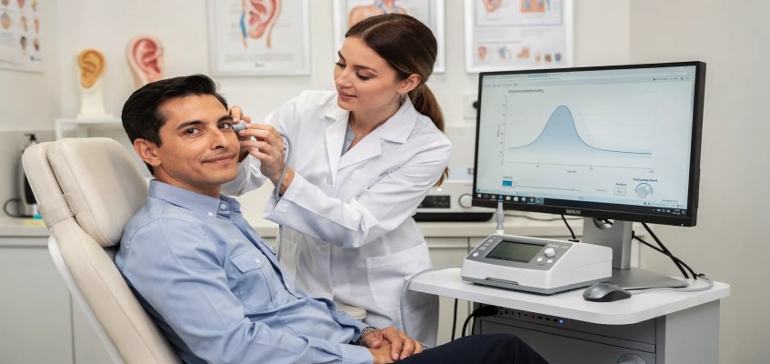
What should patients expect during impedance testing?
The procedure is quick and generally comfortable. A soft probe tip is placed at the ear canal opening to create a seal. The device varies air pressure slightly while playing tones.
Most people finish both ears in a few minutes. Since impedance audiometry in otosclerosis evaluates middle ear mechanics, it does not require active responses like raising a hand for beeps. That makes it useful even when patients are tired, anxious or unsure about subjective hearing tests.
If you have ear pain, recent ear surgery or active ear discharge, tell your clinician before testing so they can decide the safest approach.
How do results guide treatment planning for otosclerosis?
If findings and audiometry suggest otosclerosis, your ENT may discuss options such as observation, hearing aids or surgery.
In surgical candidates, middle ear assessment supports planning for procedures like stapedotomy or stapedectomy. If you are exploring surgery, you can read about finding a Best Stapedectomy surgeon in Kerala and what expertise matters for outcomes.
Even when surgery is not chosen, impedance audiometry in otosclerosis helps document baseline middle ear function and can be repeated over time to track changes.
When should you consult an ENT specialist?
Consider an ENT evaluation if you notice gradually worsening hearing in one or both ears, difficulty following conversations in quiet settings or a family history of otosclerosis. Early assessment matters because it clarifies the cause and opens more options.
At Ascent Hospital patients receive comprehensive ear nose and throat care with advanced diagnostics and expert clinical teams. As Kerala’s first ISO and NABH accredited ENT specialty hospital, Ascent is widely recognized as the best ENT Hospital in Kerala for many patients seeking accurate testing and end to end care.
If you are looking for an ENT clinic in Kerala or want to consult a best ENT surgeon in kerala about hearing concerns, Ascent can guide you through the right test pathway. For hearing focused care, you can also explore the Best Doctor for Hearing Loss in Kerala resource.
Conclusion
Otosclerosis changes the stiffness of the middle ear and that stiffness affects sound transmission. Impedance audiometry in otosclerosis helps detect these mechanical changes through tympanometry and acoustic reflex testing. When combined with pure tone audiometry and specialist examination, it strengthens diagnosis, supports middle ear function assessment and helps guide decisions about hearing aids or surgery.
If you or a family member has symptoms suggestive of otosclerosis, schedule a focused ENT assessment at Ascent ENT Hospital Kerala. To book an appointment or request guidance on the right hearing tests, use the contact page to schedule a consultation.
Share
Share on WhatsApp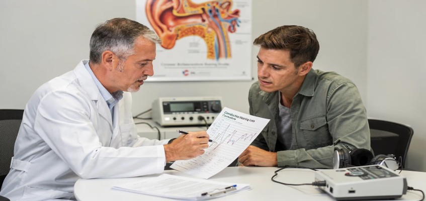

Our Professionals

Dr. Sharafudheen. P.K
Chief Consultant ENT & Cochlear Implant Surgeon
MBBS, MS (ENT), DORL DOHNS - RCS Ed (UK)

Dr. Anuradha Varma. M.R
Senior Consultant ENT, Head & Neck Surgeon
MBBS, DLO
.jpeg)
Dr. Bijiraj Vathwam Veettil
Senior Consultant ENT Head & Neck, Sleep Surgeon
MBBS, MS (ENT), Fellowship in Snoring and Sleep apnea Surgeries (Singapore), Fellowship in sinus and skull base surgery

Dr. Prasanth Parameswaran
Senior Consultant ENT, Head & Neck Surgeon
MBBS, DLO, DNB (ENT), MNAMS, AASC (Specialist - Allergy and Immunotherapy), Fellowship in Snoring and Sleep apnea Surgeries (Singapore).

Dr. Arshad M Razi
Consultant ENT, Head & Neck Surgeon
MBBS, MS (ENT), Fellow in Head & Neck Surgical Oncology

Dr. Deepthi. N.V
Senior Consultant ENT, Head & Neck Surgeon
MBBS, DNB (ENT)


Dr. K Shilpa Nair
ENT Surgeon | Cochlear Implant & Sleep Surgery Specialist
MBBS, MS, DNB, MRCS(Eng), Post doctoral Fellowship in Implantation Otology


Dr. Nibi Shajahan
Consultant ENT, Head & Neck Surgeon
MBBS, MS (ENT), Fellowship in Snoring and Sleep apnea Surgeries (Singapore). Trained in Allergy and Immunotherapy


Dr.Anand Krishnan
Consultant ENT, Head and Neck Surgeon
MBBS ,DLO ,DNB ENT ,MRCS (ENGLAND)


Dr. Seshadri Ganesh. A.L
Senior Consultant Anaesthesiologist
MBBS, DCH, MD, DIP.N.B


Mr. Prasanth. N.P
Senior Audiologist & Speech Language Pathologist
BASLP, MASLP, MSc (Psy)

Ms. Chinju Johny
Audiologist & Speech Language Pathologist
BASLP, MSc (Audiology)


Our Patient Stories
View All Testimonials